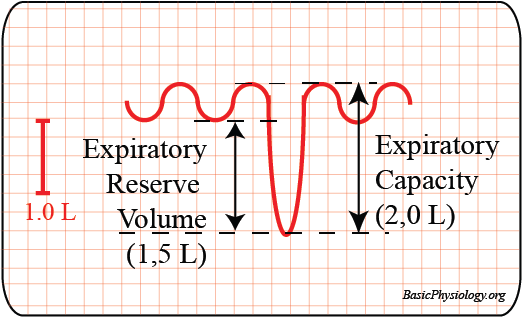
A. Introduction
1.

In the previous pages, you have seen that the air flows in and out of the lungs because of differences in the air pressure between the inside and the outside of the lungs.

2.
All these airflows are volumes of air that gets inhaled and expired.
3.
In diseases or during exercise, these volumes may increase or decrease.
4.
It would therefore be nice if we could measure these volumes. This is the task of the spirometer!
B. The Spirometer:
1.

The spirometer is a machine that measures the volume of air that is inhaled or exhaled and plots this on a paper or on a computer screen.

2.

As shown in the diagram, the spirometer actually consists of a bucket that is hanging upside-down in water. A tube connects the air trapped inside the bucket to the mouth of the patient or a volunteer (you?).

3.

When you inhale, air goes into the lungs. That air comes from the bucket. As the air volume in the bucket decreases, the bucket sinks (why?) and pulls on the string. This string pulls, through a series of wheels, the pencil that draws a curve on the graph.

4.
And when you exhale, air flows into the bucket, which will raise the bucket and the pencil trace will then lower. So, the changes in volume are reflected as changes in the amplitude of the curve.
5.

By the way, you need to close the nose of the person with a nose clip, or else some air will also go in- and out through the nose which will not be measured by the spirometer.

6.

The spirometer, as described in the diagram, is actually an old machine that is no longer really used except for teaching purposes (like here!).

7.

Currently, there are more modern machines such as the vitalograph that measure the same thing electronically.

Everything is now electronically detected so this really consists of a box with a tube attached to it. However, I like to use the old-fashioned spirometer because it is easier to explain and to understand.

Why? As the pressure in the bucket is decreased, the atmospheric pressure will push fluid into the bucket to replace this air volume so that the pressure inside the bucket remains equal to the pressure outside. So, the bucket sinks and pulls at the string!

C. Tidal Volume and Respiration Frequency
1.

When you connect a person to the spirometer or the vitalograph, his or her respiration will then be recorded and a curve drawn on the graph paper.

2.

This respiration is visible as a series of waves. From these respiration waves we can calculate the frequency and the depth of respiration.

3.

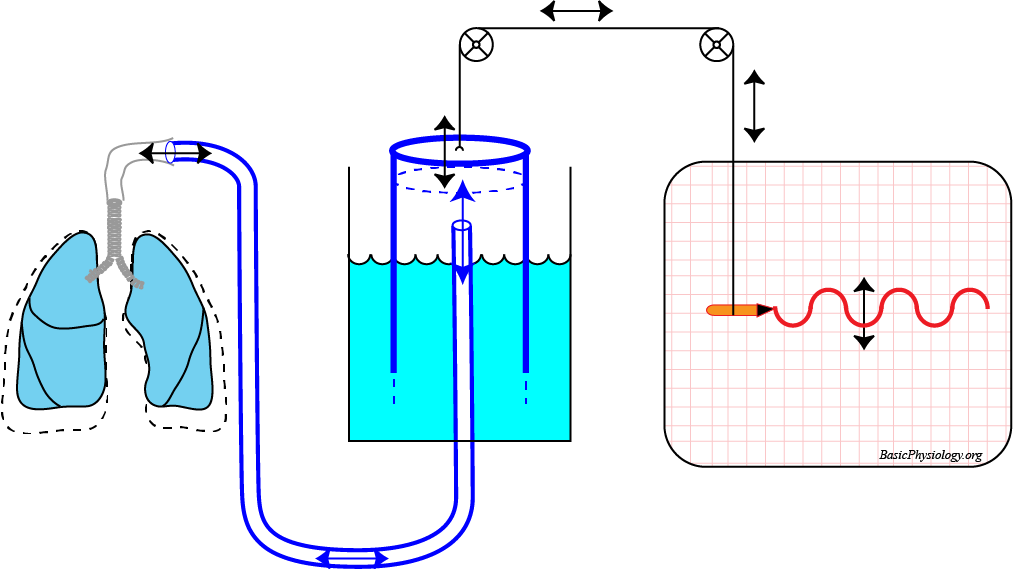
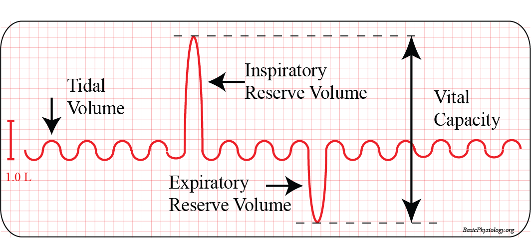
We call the volume that is in- and expired the tidal volume (= from ‘tide’; going in and out, like the sea tide at the beach).

The tidal volume is, at rest, typically about 500 ml (= half a liter!).

The frequency of respiration, again at rest, is typically 12 cpm (cycles per min).

4.

From these two values, we can easily calculate the minute ventilation (because this is calculated for one minute), also called the total ventilation.

(Remember the cardiac output = heart frequency x stroke volume? Minute Volume is the same approach but now for the lungs).

5.

So, in this situation, the minute ventilation would be:

    0.5 liter x 12 cpm = 6.0 liter/min.

Ok?

D. Tidal volume and Respiration Frequency during Exercise
1.
When the person is not at rest, but asked to exercise, then both the tidal volume and the frequency will increase. This of course will increase the minute ventilation.
2.

From this increase in the volume that is in- and expired, you can see that the lungs have a large reserve volume. These reserve volumes will be further discussed in the next section.

3.
In this case, the tidal volume increased to 2.8 L and the frequency to 23 cycles/min.
4.
The minute volume has therefore increased (from 6.0 L) to 64.4 L/min. That is more than a ten-fold increase!
E. Inspiratory and Expiratory Reserve Volumes and Capacities
1.
It is sometimes necessary to measure these reserves volumes in the lungs of a patient. Why?
2.

Therefore, when someone’s respiratory system is being tested in the hospital, you ask the patient to perform certain tests.

3.

When you ask the patient to inspire air as much as possible into his or her lungs, the curve on the graph will go up a lot. This volume, above the tidal volume, is called the Inspiratory Reserve Volume (typically about 2.0-3.0 liter).

4.

Similarly, you can ask the patient to expire as much as possible. This volume is called the Expiratory Reserve Volume(typically 1.0 – 2.0 liter). In most people, the Inspiratory Reserve volume is larger than the Expiratory Reserve volume.

5.

Confusion! Some books and some teachers don’t use the term Reserve Volume but the term Capacity. And this is slightly different!

6.

Simply put, the capacity is the reserve volume  + the tidal volume. As shown in the diagram, the Inspiratory Capacity is the Inspiratory Reserve Volume + the tidal volume. The same is true for the Expiratory Capacity.

Why? Because in several lung diseases, the reserve volumes are smaller than normal values which may affect the patient’s ability to breathe properly.

F. Vital Capacity and Total Lung Capacity
1.

This brings us to another important value; the Vital Capacity. This is very simply the sum of the Inspiratory Reserve Volume + the tidal volume + the Expiratory Reserve Volume.

2.

In the diagram, for example, the vital capacity would be:

2,500 + 0,500 + 1,500 = 4,500 ml.

3.

If your teacher insists on using capacity then Vital Capacity =

Inspiratory capacity + Expiratory capacity – Tidal volume!    (or else you would count the tidal volume twice!).

4.

Is this all? No, not quite.

Even if you exhale as much as you can, there will still be some air left in the lungs.
5.

The amount of air that you cannot exhale is called the Residual Volume (typically about 1200 ml).

6.

Finally, finally, you have the grand total of all lung volumes; the Total Lung Capacity, which is the total amount of air in the lungs; about 6 liters.

7.

In the next diagram, I have plotted all these volumes together, using color areas to indicate each type of volume. Hope this helps!

G. Function Tests:
1.

As stated above, the spirometer can measure the volumes (and capacities).

2.

It can also measure the Residual Volume with the help of some diffusion tests but this is more complicated, not often used, and is therefore not discussed here.

3.

Aside from all these volumes and capacities, it would also be useful if we could “stress-test” the lungs.

4.

This is performed, not by asking the patient how much he or she can in- or expire (= volume), but how fast he or she can breathe in or out (= speed).

5.

The most common function test in this context is the Forced Expiratory Volume in 1 second.

6.

In this test, you ask the patient to first inhale as much as possible so that the patient is at the top of the Inspiratory Volume (point ‘a’ in the diagram).

7.

Then you ask the patient to exhale as fast as possible (indicated in the diagram as the blue line, from “a” to “b”).

8.
The amount of air expired in exactly 1 second is then measured (point ‘b’ in the diagram). This volume should be, in a healthy person, at least 80% of the Vital Capacity.
9.

In this case, the patient had a vital capacity of 4.5 L and expired in one minute 3.9 L.

10.
This makes it 86.6% of the vital capacity, which is more than enough.
H. Dead Space and Alveolar Ventilation
1.

Note that not all the air that we inhale is used for the gas exchange.

2.
In fact, as we inhale, the last bit that gets sucked into the nose, mouse, pharynx, larynx, trachea and the whole bronchial tree will never be in contact with blood and will never get “exchanged”.
3.

Only the air that gets in the alveoli will be exchanged. This is the alveolar space (= volume).

4.

The amount of air that get inspired but not “exchanged” is called the dead space. It amounts usually to about 150 ml. This is essentially the total volume of the respiratory airways.

5.
Remember that the usual amount of air that gets inspired at rest, the tidal volume, is about 500 ml.
6.

In other words, the amount of fresh air that really gets into the alveoli is:

500 – 150 = 350 ml.

This is the alveolar ventilation.

Slides C.4.3. The Lungs Volumes